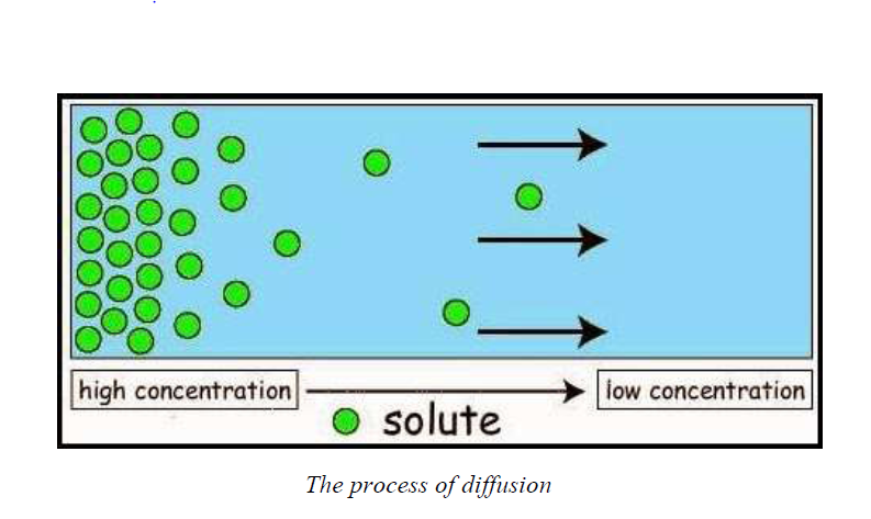
Diffusion, Osmosis and Mass- flow


Transport of Materials in Mammals, the Structure of the Mammalian Heart
The External and Internal Structures of the Mammalian Heart
Describe the external and internal structures of the mammalian heart
TRANSPORT OF MATERIALS IN MAMMALS
Mammals are the complex multicellular organisms whose bodies are made up of numerous cells and tissues. In this case, diffusion alone is not enough to insure efficient carrying out of life process. Therefore mammals have an elaborate transport system that is made up of the heart ,blood and blood vessel.
The structure of the mammalian heart
The heart is a muscular organ about the size of a closed fist that functions as the body’s circulatory pump. It takes in deoxygenated blood through the veins and delivers it to the lungs for oxygenation before pumping it into the various arteries. The heart is located in the thoracic cavity between the two lungs.
The external structure of the mammalian heart is as shown in the labelled diagram below:
The mammalian heart is broader at the top and narrower at the bottom. It is enclosed by a double layer of tough and elastic membranes called pericardium. These membranes prevent the heart from ever-expanding when beating very fast. Also the pericardium secretes a fluid which enables the membranes to move smoothly against each other.
The walls of the ventricles are thicker than those of the auricles because the ventricles pump blood a greater distance than the auricles. Auricles pump blood to the ventricles while the ventricles pump blood to the other parts of the body.
The left ventricle is thicker than the right ventricle because the right ventricle pumps blood to the lungs while the left ventricle pumps blood to the rest of the body parts.The heart consists of four chambers, right and left atria and right and left ventricles. The functions of each part and the associated structures are as follows:
- The right atrium links to the right ventricle by the tricuspid valve. This valve prevents back flow of the blood into the atrium above, when the ventricle contracts.
- The left atrium links to the left ventricle by the bicuspid valve. This valve also prevents back flow of the blood into the atrium above, when the ventricle contracts.
- Semi-lunar (pocket) valves are found in the blood vessels leaving the heart (pulmonary artery and aorta). They only allow exit of blood from the heart through these vessels following ventricular contractions.
- Ventricles have thicker muscular walls than atria. When each atrium contracts, it only needs to propel the blood a short distance into each ventricle while ventricles pump blood to distant body parts.
- The left ventricle has even thicker muscular walls than the right ventricle. The left ventricle needs a more powerful contraction to propel blood to the systemic circulation (all of the body apart from the lungs). The right ventricle propels blood to the nearby lungs. So, the contraction does not need to be so powerful.
INTERNAL STRUCTURE OF THE MAMMALIAN HEART

The heart has several valves. And these valves have flaps that ensure that blood flows in one direction only.
These valves include the following:
- The tricuspid valve; found between the right auricle and right ventricle
- The bicuspid valve: found between left auricle and left ventricle
- Semi-lunar valves which are located at the bases of the pulmonary artery and aorta to prevent blood from flowing back into the ventricles.
These valves will close if the blood flows back. The valves are held in place by tendons which prevent the flaps from turning inside out.The right and left sides of the heart are separated by septum which is a thick muscular wall which prevents mixing of oxygenated and deoxygenated blood.
The Functions of the External and Internal Parts of the Mammalian Heart
Explain the functions of the external and internal parts of the mammalian heart
Functions of parts of the mammalian heart
Part of the heart
|
Function
|
Aorta
|
The largest artery in the body; it conducts freshly oxygenated blood from the heart to the tissues.
|
Superior vena cava
|
Large vein that brings deoxygenated blood from the upper parts of the body to the right atrium
|
Inferior vena cava
|
Large vein that brings deoxygenated blood from lower regions of the body to right atrium
|
Pulmonary artery
|
Carries deoxygenated blood from the right ventricle to the lungs
|
Pulmonary vein
|
Blood vessel that carries oxygenated blood from the lungs to the left atrium
|
Right atrium
|
This chamber of the heart receives deoxygenated blood from the body (from the superior and inferior vena cava).
|
Left atrium
|
This chamber of the heart receives oxygenated blood from the lungs
|
Tricuspid valve
|
Located on the right side of the heart between the right atrium (RA)and right ventricle (RV)
|
Bicuspid valve
|
Located on the left side of the heart between the left atrium (LA) and the left ventricle (LV)
|
Right ventricle
|
The chamber of the heart that pumps deoxygenated blood to the lungs
|
Left ventricle
|
Receives blood from the left atrium and pumps it into the aorta for transport to the body cells
|
Septum
|
Divides the right and left chambers of the heart
|
The Adaptations of the Parts of the Mammalian Heart to their Functions
Explain the adaptations of the parts of the mammalian heart to their functions
The heart is adapted to carry out its functions by having the following features:
- The cardiac muscle is adapted to be highly resistant to fatigue.
- The heart has a large number of mitochondria enabling continuous supply of energy to the heart and numerous myoglobins (oxygen storing pigment).
- The presence of the cardiac muscles enables the heart to beat rhythmically.
- The pericardium which surrounds and protects the heart from physical damage.
- Pericardial fluid which prevents friction when the heart beats.
- The outer layer of the pericardium attaches to the breastbone and other structures in the chest cavity and thus helps to hold the heart in place.
- Bicuspid and tricuspid valves between atria and ventricles which prevent the backflow of blood.
- Septum which prevents the mixing of deoxygenated blood in the right and oxygenated blood in the left chambers of the heart.
- Its own blood supply for supplying nutrients and removing waste.
- The left ventricle has thick muscular wall to pump blood at a higher pressure to the distant body tissues,
- The heart is supplied with the nerves which control the rate of heartbeat depending on the body requirements.
Blood vessels
Blood vessels are intricate networks of hollow tubes that transport blood throughout the entire body. This is an essential function as blood delivers valuable nutrients to and removes wastes from our cells. Blood vessels are constructed of layers of connective tissue and muscle. The inner blood vessel layer is formed of endothelium. In capillaries and sinusoids, endothelium comprises the majority of the vessel. There are three types of blood vessels namely arteries,veins and capillaries. Each of these vessels has a different structure and function.
The Structure of Arteries, Veins and Capillaries
Describe the structure of arteries, veins and capillaries
Basic structure
- Capillaries consist of anendothelium whichis only one cell thick.
- Walls of arteries and veins consist of 3 layers.
- The inner layer consists of a thin layer of endothelial cells.
- The middle layer is made up of smooth muscle with some elastic fibres. This layer controls the diameter of the vessel and hence the amount of blood and its rate of flow.
- The outer layer is composed of connective tissue; this holds the blood vessels in place in the body.
Detailed structure
Arteries
- The walls of arteries are much thicker as it carries blood away from the heart at high pressure.
- Major arteries close to the heart also have thick layers of smooth muscle in their walls to withstand the increases in pressure as the heart pumps.
- The walls also have a large proportion of elastic fibres in both the inner and middle layers – this allows for the arteries to stretch according to the increases in volume of blood. As the heart relaxes the artery walls return to their original position, hence pushing the blood along – maintaining a constant flow in one direction.
- Arteries are near the surface of the skin; the changes in the arteries diameter can be felt as a pulse.
Veins
- The walls of veins are thinner than the walls of arteries, as the blood they receive from the capillaries is at a much lower pressure.
- The walls have fewer elastic fibres and the lumen is wider (to allow for easier blood flow).
- Veins have two mechanisms for keeping the blood flow constant and in one direction. Firstly, many veins are close to muscles, hence when the muscles contract they compress the walls of the vein – pumping blood forwards. Veins also have valves which are spaced along regular intervals in veins. They work much like one-way swinging doors – as the blood is forced through the valve opens. However, once the pressure drops and the blood flow decreases, the valve shuts – preventing back flow of blood.
Capillaries
- They are extremely, tiny microscopic vessels that bring blood into close contact with the tissues, for the exchange of chemical substances between cells and the bloodstream.
- The one cell thick endothelial layer is a continuation of the lumen arteries and veins.
- Diffusion is a relatively slow process and hence the structure of capillaries is suited to slowing down the flow of blood.
- In order to maximize the exchange of substances between the blood and cells, capillaries have thin walls (for more efficient diffusion) a small lumen (that forces blood cells to pass through in single file, slowing down the rate of flow and maximizing their exposed surface area).
- They form an expansive blood flow network, such that no cells are far from blood supply

How different blood vessels are adapted for their function
Blood vessel
|
Function
|
Adaptation
|
Artery
|
Carries blood away from heart at high pressure
|
Thick, elastic, muscular walls to withstand pressure and to exert force (pulse)
|
Vein
|
Returns low-pressure blood to heart
|
Large diameter to offer least flow resistance. Valves to prevent back flow.
|
Capillary
|
Allows exchange of materials between blood and tissues
|
Thin, permeable walls
|

Differences between arteries, veins and capillaries
Arteries
|
Veins
|
Capillaries
|
All arteries carry blood away from the heart
|
All veins carry blood towards the heart
|
Capillaries carry blood from arteries to the veins
|
With the exception of the pulmonary artery, all arteries carry oxygenated blood
|
With the exception of the pulmonary vein, all veins carry deoxygenated blood
|
Blood slowly loses its oxygen
|
They carry blood which is usually rich in digested food materials
|
Except for the hepatic portal vein, they carry blood which usually has little digested food materials
|
Blood slowly loses its food
|
Have relatively narrower lumens (see diagrams above)
|
Have relatively wide lumens (see diagrams above)
|
Have relatively narrow lumens (see diagrams above)
|
Have relatively a thick layer of muscles and elastic fibres
|
Have relatively a thin layer of muscles and elastic fibres
|
They do not have muscles and elastic fibres
|
They have thick outer walls
|
They have thin outer walls
|
Walls are only one cell thick
|
They carry blood at high pressure
|
They carry blood at low pressure
|
Pressure gradually falls as blood flows from arteries to veins
|
Do not have valves (except for the semi-lunar valves of the pulmonary artery and the aorta)
|
Have valves throughout the main veins of the body to prevent the back flow of blood.
|
Have no valves
|
Have bright red blood (because it is rich in oxygen)
|
Brown-red blood
|
Brown-red blood
|
Located deep in the to body surface
|
Located near to body surface
|
Capillaries are found inside all tissues
|
Walls are not permeable
|
Walls are not permeable
|
Walls are permeable
|
Blood flows in pulses
|
No pulse
|
Pulse gradually disappears
|
Simple Experiments to Determine Pulse Rates in Human Being
Carry out simple experiments to determine pulse rates in human being
Carry out simple experiments to determine pulse rates in human being
The Blood
The Major Components of the Blood
List the major components of the blood
Blood is the red fluid that circulates in our blood vessels. The average human body containsabout 4 to 5 litres of blood. Blood is classified as a connective tissue and consists of two maincomponents:
- Plasma which is a clear extracellular fluid.
- The solid component, which are made up of the blood cells and platelets
The solid component is made up of blood cells except for the platelets, which are tiny fragmentsof bone marrow cells.
The solid component consists of blood cells (corpuscles) which include:
- Erythrocytes, also known as red blood cells (RBCs)
- Leukocytes, also known as white blood cells (WBCs)
- Platelets, also known as thrombocytes
Red blood cells, most white blood cells, and platelets are produced in the bone marrow, the soft fatty tissue inside bone cavities. The white blood cells (lymphocytes) are also produced in the lymph nodes and spleen, and in the thymus gland.
Within the bone marrow, all blood cells originate from a single type of unspecialized cell calleda stem cell. When a stem cell divides, it first becomes an immature red blood cell, white bloodcell, or platelet-producing cell. The immature cell then divides, matures further, and ultimatelybecomes a mature red blood cell, white blood cell, or platelet.
Blood cells

By volume, the plasma constitutes about 55% of whole blood, and red blood cells, platelets and white blood cells about 45%.

The Function of Major Blood Components
Explain the function of major blood components
Red blood cells
Red blood cells (RBCs) have two main functions:
- To pick up oxygen from the lungs and deliver it to tissues elsewhere.
- To pick up carbon dioxide from other tissues and unload it in the lungs.
Erythrocytes transport oxygen in the blood through the red pigment called haemoglobin.Haemoglobin contains iron and proteins joined to greatly increase the oxygen carrying capacity of erythrocytes. The high surface area to volume ratio of erythrocytes allows oxygen to be easily transferred into the cells in the lungs and out of the cells in the capillaries of the systemic tissues.Erythrocytes are produced inside red bone marrow from stem cells at the astonishing rate of about 2 million cells every second.
White blood cells
Although the white blood cells accounts for only about 1% of the blood, they play a very important role in the body. Their main function is to protect the body against disease pathogens.There are two of white blood cells, each of which plays a specific role in protection of the body against illness and disease.
- Phagocytes: Engulf and digest invading bacteria and viruses (pathogens). It is the body’s main defence against germs (microbes).
- Lymphocytes: produce antibodies which neutralize antigens from bacteria or viruses. They kill microbes or make them clump together, to be removed in the lymph glands.
White blood cells are produced in the yellow marrow of the bone, spleen, thymus and lymphatic system.
Platelets
Platelets are small fragments of bone marrow cells and are therefore not really classified as cells themselves. Platelets have the following functions:
- Secrete vasoconstrictors which constrict blood vessels, causing vascular spasms in broken blood vessels.
- Form temporary platelet plugs to stop bleeding.
- Secrete procoagulants (clotting factors) to promote blood clotting.
- Dissolve blood clots when they are no longer needed.
- Digest and destroy bacteria.
- Secrete chemicals that attract neutrophils and monocytes to sites of inflammation.
- Secrete growth factors to maintain the linings of blood vessels.
In general, the blood platelets functions in healing of the wounds when the skin gets broken. This is achieved by clumping together of the platelets to form a network of mesh, hence bleeding is stopped.
Plasma
Plasma is the non-cellular or liquid portion of the blood. Plasma is a mixture of water, proteins, and dissolved substances. Around 90% of plasma is made of water, although the exact percentage varies depending upon the hydration levels of the individual. Blood plasma has the following functions:
- Plasma serves as a transport medium for delivering nutrients to the cells of the various organs of the body.
- It transports waste products derived from cellular metabolism to the kidneys, liver, and lungs for excretion.
- It fights infections since it contains antibodies.
- It is also a transport system for blood cells, and it plays a critical role in maintaining normal blood pressure.
- Plasma helps to distribute heat throughout the body and to maintain homeostasis, or biological stability, including acid-base balance in the blood and body
- It carries and transports some hormones.
The Effects of HIV on White Blood Cells
Explain the effects of HIV on white blood cells
The HIVs in the blood of a HIV-positive person attack the white blood cells (lymphocytes). The viruses reproduce and increase in number within the lymphocytes. Then the lymphocytes burst and release more viruses in the bloodstream. The released viruses attack more, new white cells.The attack continues in that cycle until many white cells are destroyed. Because there are only a few white cells left to fight against pathogens, the body immunity gets low. Once the immunity is lowered, the body is often attacked by diseases and a person suffers from AIDS.
Blood Groups and Blood Transfusion

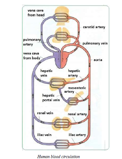
Blood Circulation
The Lymphatics System

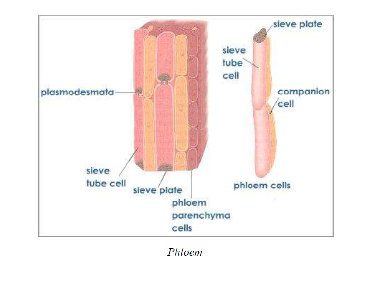
Transport of Material in Plants in Plants, the Vascular System


...
[
[


No comments:
Post a Comment